Mirabiliss Poliklinika, Niš - Website logo

Mirabiliss Polyclinic - Instagram
Mirabiliss Poliklinika, Niš - Facebook


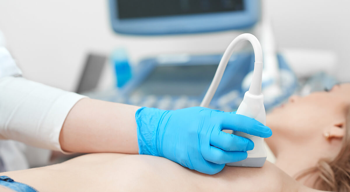
    Ultrasound examination of the breast

    Thanks to advances in technology, sophisticated equipment and software have been created that enable precise breast examinations with ultrasound devices.

    Ultrasound is especially important in detecting benign changes in the breasts and distinguishing them from malignant ones, which is often not possible with mammography, especially in breasts with dense glandular tissue.

    In women younger than 40, but also those older who have dense glandular tissue, ultrasound examination of the breast is the first choice.

    Ultrasound works on the principle of harmless, high-frequency sound waves that are converted into an image with the help of a suitable computer program. Namely, sound waves pass through the breast tissue, bounce off various structures in the breast and return back to the probe, which, with the help of appropriate software, enables the monitoring of the image on the monitor. As the tissues are of different structure, so are the sound waves of different characteristics, on the basis of which they can differ from each other.

    This method of examination is not aggressive, ie. it does not damage tissue or cause radiation. The most common application is to examine a certain area in the breast where there is a problem. A palpable nodule and / or a shadow or thickening in the breast detected by mammography can be further analyzed by ultrasound.

    The particular benefit of ultrasound is in distinguishing between fluid-filled cysts from solid tissue constructions.  Recent studies show that in the diagnosis of breast cancer in women younger than 45, ultrasound is a more accurate method than mammography.

    Mirabiliss Polyclinic - Nis - Ultrasound examination of the breast 04
    Mirabiliss Polyclinic - Nis - Ultrasound examination of the breast 08

    Due to the fact that there is no radiation, ultrasound is an ideal method of breast examination in pregnant women. Checking breast implants is the next big benefit of ultrasound. Inflammatory conditions in the breast, such as the formation of inflammatory pockets or abscesses, can be identified and monitored by ultrasound.

    Thick and edematous skin can be a sign of inflammatory breast cancer. Ultrasound can identify malignant growths inside the breast that cause skin thickening. Ultrasound can also determine exactly how far the process has spread in the breast, that is, how much it has penetrated into healthy tissue.

    Ultrasound examination is performed in an outpatient setting. The examination is performed on an appropriate examination table, and the patient should be taken off clothes to the waist so that the breasts, axillary pits and the area above the collarbones can be examined. The examination itself is performed in a supine position on the back or side. A gel is applied to the skin, which is a necessary enhancer of ultrasonic wave transmission. The examination usually lasts about 30 minutes, and it is best to do it in the first ten days of the menstrual cycle, when the breasts are less painful and tense, and the influence of female sex hormones is not as pronounced as in the premenstrual period.

    Ultrasound examination may show normal breast tissue or reveal benign changes such as simple cysts that do not produce symptoms of the disease and that do not require any further treatment other than regular monitoring. Such findings are considered normal findings. Pathological findings can be classified into one of the following groups: benign fibrous nodules, more complex cysts, suspicious lesions, or lesions highly suggestive of cancer. Patients with such findings are referred for further diagnosis and treatment.